New mouse model offers key insights into gut colonization by antibiotic-resistant bacteria
MSU researchers identified critical genes that help Enterobacter hormaechei thrive in the gut, paving the way for new infection-fighting strategies.
The spread of antibiotic-resistant bacteria is becoming a serious, global public health problem. Some bacteria are developing resistance to carbapenems, the most powerful antibiotics that we have available, leaving healthcare providers with limited treatment options and leading to terrible consequences.
One such bacterium, Enterobacter hormaechei, is responsible for causing a wide variety of severe infections, including life-threatening septic shock in newborns and immunocompromised adults. However, E. hormaechei doesn’t always pose a threat. It can coexist harmlessly in the gut, becoming a problem only when something disturbs the balance between them and the beneficial microbes that also live there, allowing the pathogen to multiply and enter the bloodstream.
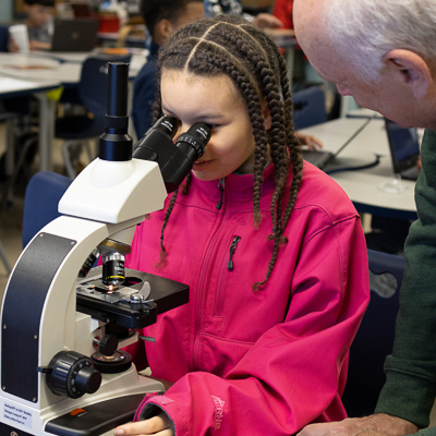
“Once they start causing infection, they can become really hard to treat. That’s why we’re studying them,” said Beth Ottosen, a doctoral student in MSU’s department of Microbiology, Genetics, & Immunology, or MGI. “The research that I do is looking at how, specifically, Enterobacter is able to survive in our body and cause these infections, but also how it’s able to live in the gut, since that’s such a big reservoir for these infections.”

Ottosen, who works in the lab of MGI department chair and Rudolph Hugh Endowed Chair Victor DiRita, hopes that understanding the mechanisms behind how E. hormaechei colonizes the human gut will help identify strategies to combat these dangerous infections.
In a recent paper published in mBio, Ottosen and Ritam Sinha, a research scientist also working in the DiRita lab, developed a new neonatal mouse model to better study infections in human infants. Using that model, they found evidence that E. hormaechei feeds on mucus in the intestines.
To learn more about the relationship between E. hormaechei and intestinal mucus, the researchers examined an enzyme called pyruvate dehydrogenase complex, or PDH. This enzyme helps cells get energy from food and is already known to be important in the growth of several other gut bacteria.
Using genetic engineering, they created a version of E. hormaechei that couldn’t produce PDH, then tested how well it could grow on the various sugars found in the intestinal mucus. As Ottosen explained, “Mucus is a glycoprotein. It’s a protein decorated with a lot of sugars.” The mutant bacteria couldn’t grow on any of the sugars except for one called GlcNAc, or N-acetyl-D-glucosamine. Next, they studied the genes that help the bacteria metabolize GlcNAc and created another version that couldn’t use GlcNAc as food.
“We found that a mutant in either of those pathways doesn’t do so great; it colonizes about half as well as wild type,” Ottosen said. “But a mutant in both of those pathways at once can’t colonize your gut at all, and it cannot grow on mucin.”
These results show that these two genes are important for E. hormaechei to survive and thrive in the gut. This makes them good targets for developing new treatments against infections caused by these drug-resistant bacteria.
“This new model of E. hormaechei colonization and growth in the gut of neonates will let us explore the biology and pathogenicity of this important microbe,” said DiRita, “We’re interested in fundamental questions such as the role that breast-feeding might play in protecting infants from sepsis, and whether we can intervene to reduce colonization by the bug in the first place”.