Research
Beyond Snapshots: Visualizing the Dynamic Life of Immune Cells
The immune system is a highly orchestrated network where cell-to-cell communication and spatial localization determine the outcome of an immune response. Our lab utilizes Intravital Microscopy to observe immune cells in their native environment within a living host.
A hallmark of our research is the use of Three-Photon (3P) microscopy, which enables high-resolution visualization of deep tissues that remain inaccessible to conventional Two-Photon (2P) microscopy. By capturing real-time interactions, we aim to uncover novel molecular mechanisms of immunity—focusing on how the unique microenvironments of lymph nodes and the spleen coordinate systemic immune responses.
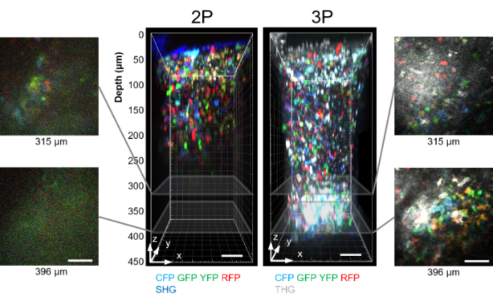
Imaging depth comparison of 2P and 3P microscopy in the same popliteal lymph node of Cγ1Cre-Confetti mouse in vivo, in which germinal center B cells express one or two of four different fluorescent
proteins (CFP/GFP/YFP/RFP).
Source: Choe et al., Nature Immunology (2022)
1. Decoding Immune Orchestration in the Lymph Node
Lymph nodes serve as critical surveillance hubs, filtering lymph-borne antigens and pathogens to initiate adaptive immunity. Understanding immune cell behavior in lymph nodes is fundamental to advancing vaccine strategies and immunotherapy. By leveraging our pioneering intravital 3P microscopy, we investigate how T and B cells migrate and interact within deep lymph node regions that were previously "blind spots." Our studies span steady-state homeostasis, infectious conditions, and malignancy (such as lymphoma) to decipher the spatial logic of immune activation.
Video: Migration of CD8 (red) and CD4 (green) T cells at different depth (Z) of lymph node.
Source: Choe et al., Nature Immunology (2022)
2. Splenic Lymphocyte Trafficking
Despite its massive role in filtering blood, the spleen remains one of the least understood organs in terms of real-time lymphocyte trafficking due to its unique physiological landscape and significant imaging challenges. We investigate the fundamental rules of how T and B cells enter and exit the spleen and how these patterns are reprogrammed during infection.
To address the extreme motion artifacts inherent in splenic imaging, we utilize a uniquely designed, switchable microscope system. While using the up-right mode for standard lymph node imaging, the system can be converted into an inverted mode specifically for the spleen. This inverted configuration provides superior mechanical stability and minimizes tissue movement, enabling unprecedented high-resolution stabilization. Additionally, combined with our implanted imaging window, we track the same splenic niches over multiple days, providing a continuous, longitudinal view of immune response progression.

Intravital 3P microscopy of mouse spleen. Alexa-Fluor-647+ blood vessels (red) and third-harmonic generation (THG, green) signal
were imaged in spleen of adult mouse by 1650 nm 3PM. Red pulp (RP), margianl zone
(MZ), white pulp (WP).
Source: Choe et al., Nature Immunology (2022)